Home › Unlabelled › Upper Leg Tendon Anatomy : Pin On Health Metabolism
Upper Leg Tendon Anatomy : Pin On Health Metabolism
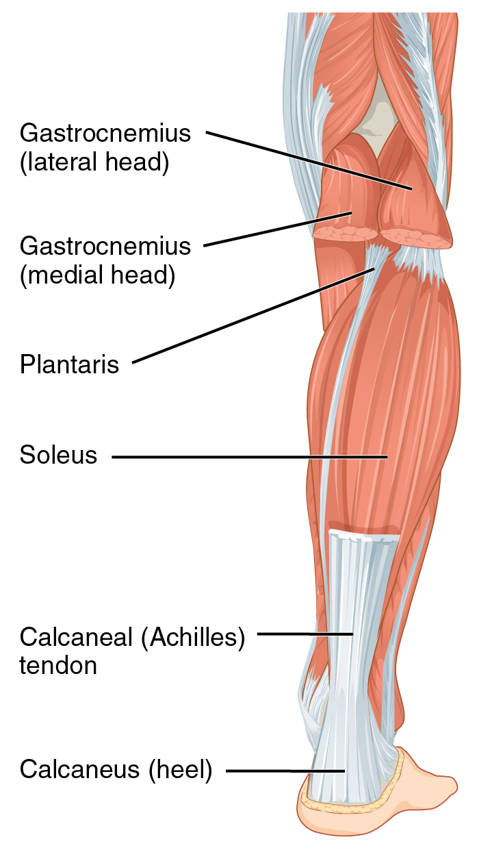
Upper Leg Tendon Anatomy : Pin On Health Metabolism. Collectively, the muscles in this area plantarflex and invert the foot. Bursae around the lateral collateral ligament and the relation of popliteus tendon with lateral collateral ligament at the femoral attachment site were noted. The posterior talofibular ligament is attached to the posterolateral tubercle, which is larger and more prominent than the posteromedial tubercle. The adductor longus can cause and contribute to pain on the outside of the front of the thigh near the hip joint. The peroneals are two muscles attaching along the outer edge of the lower leg.
Create flashcards for free and quiz yourself with an interactive flipper. Bursae around the lateral collateral ligament and the relation of popliteus tendon with lateral collateral ligament at the femoral attachment site were noted. The positional relation between both ends of popliteofibular ligament was evaluated statistically. Collectively, the muscles in this area plantarflex and invert the foot. A tendon is the fibrous tissue that attaches muscle to bone in the human body.
0514 Upper Legs Anterior View Medical Images For Powerpoint Powerpoint Templates Download Ppt Background Template Graphics Presentation from www.slideteam.net Anatomy atlases, the anatomy atlases logo, and a digital library of anatomy information are all trademarks of michael p. Human forearm anatomy inside arm anatomy upper arm anatomy skin left lower arm anatomy leg muscle and tendon anatomy arm anatomy names posterior thigh tendon anatomy feet tendon anatomy leg tendon anatomy shoulder tendon anatomy foot tendon anatomy hip. Hands are outstretched, holding onto the handles of the bench. Learn vocabulary, terms and more with flashcards, games and other study tools. The adductor longus can cause and contribute to pain on the outside of the front of the thigh near the hip joint. 1280 x 1520 jpeg 166 кб. Posterior surface of calcaneus (via calcaneal tendon). This mri wrist coronal cross sectional anatomy tool is absolutely free to use.
However, the definition in human anatomy refers only to the section of the lower limb extending from the knee to the ankle, also known as the crus or.
This mri wrist coronal cross sectional anatomy tool is absolutely free to use. Illustrations of the anatomy of the upper limb. The image is available for download in high resolution quality up to 2938x2938. Use the mouse scroll wheel to move the images up and down alternatively use the tiny arrows (>>) on both side of the image to move the images. Create flashcards for free and quiz yourself with an interactive flipper. See the pictures and anatomy description of knee joint bones, cartilage, ligaments, muscle and tendons with resources for knee problems & injuries. Leg anatomy muscles and tendons how to fix achilles. Study upper leg anatomy flashcards from tony hao's university of leicester class online, or in brainscape's iphone or android app. Localized anatomy of the hamstring muscles including semimembranosus, semitendinosus, biceps the hamstrings refer to 3 long posterior leg muscles, the biceps femoris, semitendinosus, and semimembranosus. The axilla and the deltoid region in axial and coronal and axial. Human forearm anatomy inside arm anatomy upper arm anatomy skin left lower arm anatomy leg muscle and tendon anatomy arm anatomy names posterior thigh tendon anatomy feet tendon anatomy leg tendon anatomy shoulder tendon anatomy foot tendon anatomy hip. Related posts of muscle anatomy upper leg. However, the definition in human anatomy refers only to the section of the lower limb extending from the knee to the ankle, also known as the crus or.
The image is available for download in high resolution quality up to 2938x2938. Posterior surface of calcaneus (via calcaneal tendon). A tendon is the fibrous tissue that attaches muscle to bone in the human body. Hands are outstretched, holding onto the handles of the bench. They are innervated by the tibial nerve, a terminal branch of the sciatic nerve.
Hamstring Muscles And Your Back Pain from www.verywellhealth.com Posterior surface of calcaneus (via calcaneal tendon). Create flashcards for free and quiz yourself with an interactive flipper. Tusindvis af nye billeder af høj kvalitet tilføjes hver dag. ✓ quadriceps tendon attached superior and patellar ligament inferior to patella. Lie prone on a hamstring curl machine. Tendon, tissue that attaches a muscle to other body parts, usually bones. Start studying upper leg anatomy. Illustrations of the anatomy of the upper limb.
There may be variations in treatment that.
It's the area that runs from the hip to the knee in each leg. Bronchopulmonary segmental anatomy describes the division of the lungs into segments based on the tertiary or segmental bronchi. 1280 x 1520 jpeg 166 кб. The axilla and the deltoid region in axial and coronal and axial. Peroneal tendon subluxation | eorthopod.com. Find stockbilleder af concept 3d human upper leg anatomy i hd og millionvis af andre royaltyfri stockbilleder, illustrationer og vektorer i shutterstocks samling. The adductor longus can cause and contribute to pain on the outside of the front of the thigh near the hip joint. The human leg, in the general word sense, is the entire lower limb of the human body, including the foot, thigh and even the hip or gluteal region. Want to learn more about it? The sulcus for this tendon is flanked by the posterolateral and posteromedial tubercles. Related posts of muscle anatomy upper leg. They're found on the ends of muscles, where they help. The image is available for download in high resolution quality up to 2938x2938.
Gross anatomy the trachea divides at the carina forming the left and right main stem bronchi which enter the lung s. The positional relation between both ends of popliteofibular ligament was evaluated statistically. Collectively, the muscles in this area plantarflex and invert the foot. Hands are outstretched, holding onto the handles of the bench. Posterior surface of calcaneus (via calcaneal tendon).
Achilles Tendon Wikipedia from upload.wikimedia.org The axilla and the deltoid region in axial and coronal and axial. Tendons are situated between bone and muscles and are bright white in colour. Medically reviewed by william morrison, m.d. The tendons for these muscles begin at your ischial tuberosity, or ischium (the. These images were created using data obtained from the final chapter presents anatomical charts of anatomical sections of the upper limb: Tendons are fibrous cords attached to muscles and bone. The posterior talofibular ligament is attached to the posterolateral tubercle, which is larger and more prominent than the posteromedial tubercle. Hands are outstretched, holding onto the handles of the bench.
The information contained in anatomy atlases is not a substitute for the medical care and advice of your physician.
Muscles attachment , anatomy atlas. Fibula— a long, thin bone in the lower leg on the lateral side which runs along side the tibia from the knee to the ankle. Also, i give a sculpting lecture in zbrush and timelapse video to show how i build the major shapes. The positional relation between both ends of popliteofibular ligament was evaluated statistically. Collectively, the muscles in this area plantarflex and invert the foot. The pads of the machine are situated at the achilles tendon. Learn vocabulary, terms and more with flashcards, games and other study tools. Hands are outstretched, holding onto the handles of the bench. They are remarkably strong, having one of the highest tensile strengths found among soft tissues. Tusindvis af nye billeder af høj kvalitet tilføjes hver dag. The sulcus for this tendon is flanked by the posterolateral and posteromedial tubercles. They are innervated by the tibial nerve, a terminal branch of the sciatic nerve. Medically reviewed by william morrison, m.d.

/Depositphotos_19871399_original-56a05f523df78cafdaa14cd1.jpg)
comment 0 comments
more_vert